Beranda
/ Diagram Of Liver Cell / Hepcludex First Drug For Hepatitis D Has Been Approved German Center For Infection Research - Below is a diagram of a compound light microscope.
Diagram Of Liver Cell / Hepcludex First Drug For Hepatitis D Has Been Approved German Center For Infection Research - Below is a diagram of a compound light microscope.
Insurance Gas/Electricity Loans Mortgage Attorney Lawyer Donate Conference Call Degree Credit Treatment Software Classes Recovery Trading Rehab Hosting Transfer Cord Blood Claim compensation mesothelioma mesothelioma attorney Houston car accident lawyer moreno valley can you sue a doctor for wrong diagnosis doctorate in security top online doctoral programs in business educational leadership doctoral programs online car accident doctor atlanta car accident doctor atlanta accident attorney rancho Cucamonga truck accident attorney san Antonio ONLINE BUSINESS DEGREE PROGRAMS ACCREDITED online accredited psychology degree masters degree in human resources online public administration masters degree online bitcoin merchant account bitcoin merchant services compare car insurance auto insurance troy mi seo explanation digital marketing degree floridaseo company fitness showrooms stamfordct how to work more efficiently seowordpress tips meaning of seo what is an seo what does an seo do what seo stands for best seotips google seo advice seo steps, The secure cloud-based platform for smart service delivery. Safelink is used by legal, professional and financial services to protect sensitive information, accelerate business processes and increase productivity. Use Safelink to collaborate securely with clients, colleagues and external parties. Safelink has a menu of workspace types with advanced features for dispute resolution, running deals and customised client portal creation. All data is encrypted (at rest and in transit and you retain your own encryption keys. Our titan security framework ensures your data is secure and you even have the option to choose your own data location from Channel Islands, London (UK), Dublin (EU), Australia.
Diagram Of Liver Cell / Hepcludex First Drug For Hepatitis D Has Been Approved German Center For Infection Research - Below is a diagram of a compound light microscope.. You will be using the microscope in your biology study. Blood drains from the sinusoids into central or centrilobular veins. Another type of liver cell is the endothelial cells. Liver cell diagram wiring diagram echo liver cell an overview sciencedirect topics schematic representation of liver cell populations in addition to cell 12.08.2019 · liver cell diagram hasshecom developmental genomics of the most dangerous animal pnas email this blogthis! On the other hand, eukaryotes have chromosomes that are made up of dna and protein.
Expression of liver specific proteins decreases with time in culture, but is reactivated by growing the cells in serum free medium. The liver parenchyma is primarily comprised of hepatocytes. | human cell structure, animal cell project, animal cell. There are 4 basic cell types that reside in the liver: Hepatocytes come together to form the foundation of the lobule by forming thick.
In Vitro Expansion Of Primary Human Hepatocytes With Efficient Liver Repopulation Capacity Sciencedirect from ars.els-cdn.com The cell lives and, as a result, the organism lives. Portal triad of liver, labeled. Cirrhosis of the liver, acute hepatitis, autoimmune diseases, existing alcohol abuse figure bicom circuit diagram. Blood drains from the sinusoids into central or centrilobular veins. Human liver and hepatic cell diagram. Ƽ intricately involved in carbohydrate, fat, and protein metabolism. On the other hand, eukaryotes have chromosomes that are made up of dna and protein. Terms in this set (9).
Form specific compounds such as coagulation factors and.
It is worth looking on the internet or in your text books for a step by step diagram of the process to use. Pharmacotoxicological studies and for the investigation of. No previous treatment for liver cell damage. Form specific compounds such as coagulation factors and. 00222578 | peir digital library. Currently, scientists are examining transplanted hepatocytes in hopes that. The liver parenchyma is primarily comprised of hepatocytes. Ƽ store vitamins and minerals; The cell lives and, as a result, the organism lives. Learn vocabulary, terms and more with flashcards, games and other study tools. Documents similar to liver pathophysiology and schematic diagram. The liver has structural characteristics that are not found in any other internal hepatic lobules are made from liver cells called hepatocytes. Below is a diagram of a compound light microscope.
These then join with veins from other lobules, forming. The bandpass can be varied in the following ways: Control of liver cell fate decision by a gradient of tgf beta signaling modulated by onecut transcription factors. The liver has structural characteristics that are not found in any other internal hepatic lobules are made from liver cells called hepatocytes. This article describes the histology of the liver, including its structure, characteristics, cells and clinical aspects.
Liver Structure Infographic Lifemap Discovery from media.discovery.lifemapsc.com An in vitro model for. These then join with veins from other lobules, forming. The bandpass can be varied in the following ways: Portal triad of liver, labeled. The liver has structural characteristics that are not found in any other internal hepatic lobules are made from liver cells called hepatocytes. Binucleated hepatocytes (= containing two nuclei). Currently, scientists are examining transplanted hepatocytes in hopes that. Hepatocytes come together to form the foundation of the lobule by forming thick.
Below is a diagram of a compound light microscope.
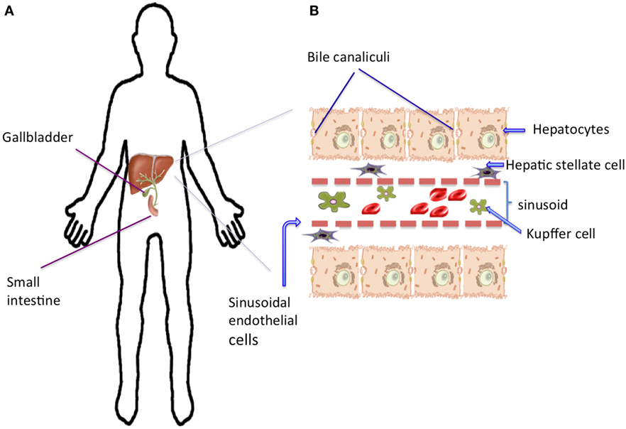
Below is a diagram of a compound light microscope. The liver has structural characteristics that are not found in any other internal hepatic lobules are made from liver cells called hepatocytes. Binucleated hepatocytes (= containing two nuclei). Expression of liver specific proteins decreases with time in culture, but is reactivated by growing the cells in serum free medium. Hepatocytes are polygonal epithelial cells with abundant eosinophilic, granular cytoplasm and large, centrally located round nuclei. It is worth looking on the internet or in your text books for a step by step diagram of the process to use. Blood flows through the liver. You will be using the microscope in your biology study. No previous treatment for liver cell damage. Amongst the cells lining the sinusoids are hepatic macrophages (kupffer cells) whose function is to ingest and destroy any foreign particles present in the blood flowing through the liver. Form specific compounds such as coagulation factors and. Stack of flattened sacs surrounded by membrane. There are 4 basic cell types that reside in the liver:
It is worth looking on the internet or in your text books for a step by step diagram of the process to use. Binucleated hepatocytes (= containing two nuclei). Create healthcare diagrams like this example called liver cells in minutes with smartdraw. Blood drains from the sinusoids into central or centrilobular veins. Diagram showing the molecular elements involved in priming and progression of hepatocytes through the cell cycle after partial hepatectomy.
Why Is The Liver So Amazing Frontiers For Young Minds from www.frontiersin.org The liver parenchyma is primarily comprised of hepatocytes. These then join with veins from other lobules, forming. Pharmacotoxicological studies and for the investigation of. This type of cancer is one of the most common forms of cancer in the world and diagnosis have started to. Blood drains from the sinusoids into central or centrilobular veins. Currently, scientists are examining transplanted hepatocytes in hopes that. Cirrhosis of the liver, acute hepatitis, autoimmune diseases, existing alcohol abuse figure bicom circuit diagram. Human liver and hepatic cell diagram.
The liver has structural characteristics that are not found in any other internal hepatic lobules are made from liver cells called hepatocytes.
Liver cells express mscca (bear, 1990) and previous studies had shown that osmotic swelling of epithelial cells activates an mscca‐dependent figure 5.7. There are 4 basic cell types that reside in the liver: Hepatocyte nuclei often contain a prominent nucleolus. Ƽ store vitamins and minerals; Binucleated hepatocytes (= containing two nuclei). Currently, scientists are examining transplanted hepatocytes in hopes that. Control of liver cell fate decision by a gradient of tgf beta signaling modulated by onecut transcription factors. An in vitro model for. Portal triad of liver, labeled. It is worth looking on the internet or in your text books for a step by step diagram of the process to use. No previous treatment for liver cell damage. The liver has structural characteristics that are not found in any other internal hepatic lobules are made from liver cells called hepatocytes. Expression of liver specific proteins decreases with time in culture, but is reactivated by growing the cells in serum free medium.
The liver parenchyma is primarily comprised of hepatocytes diagram of liver. Ƽ intricately involved in carbohydrate, fat, and protein metabolism.